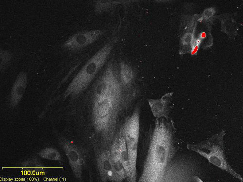
2PA Microscopy

The imaging tool is an Olympus IX70 inverted microscope with 10, 20, 40 and 60X objectives. This microscope can work in several modes: For example, Nomarski (differential interference contrast), Hg lamp excited fluorescence or as a confocal fluorescence microscope with a Fluoview scan-unit with a 488 nm Ar+ laser as an excitation source. The confocal microscope can make 3D images by letting the collected light pass through a pinhole that selects a specific plane of observation.
Layers of images can then be obtained by moving the specimen distance with respect to the microscope objective. Capture of images can be done either with the confocal software or by a high resolution video mode CCD camera (QImaging).
The confocal microscope has been modified to work as a two-photon microscope by coupling a Ti:Sapphire laser into the system.
The Ti:Sapphire is a Mira 900 (Coherent) pumped by a 10 W Verdi Nd:Vanadate laser. The Mira is a mode locked laser capable of producing light pulses of duration of ~120 fs FWHM @ 76 MHz repetition rate in a wavelength range from 700-1000 nm. The average power is > 1W.
The wavelength range and pulse duration makes this laser an excellent excitation source for the nonlinear two-photon process exploited in a 2-photon microscope. Both wavelength and pulse-duration of the laser is continuously monitored in real time.
Current efforts are to improve dyes for two-photon imaging. This includes: two-photon cross section (measure for the effectivity of the two-photon absorption process), Excitation wavelength of interest, photo-stability, and fluorescence quantum yields.
For studying higher order nonlinear absorption processes The Mira can be coupled into an Optical Parametric Oscillator capable of producing fs pulsed laser radiation from 1050 – 1400 nm (with a KTP crystal in the OPO cavity) and 1350 – 1600 nm (PP crystal in the OPO cavity) at the repetition rate of the Mira. The wavelength tuning of the OPO is obtained by tuning the pump wavelength. The OPO radiation can be coupled into the microscope

